Faili:Schistosoma haematobium egg 4843 lores.jpg

Hakuna saizi kubwa zaidi.
Schistosoma_haematobium_egg_4843_lores.jpg (piseli 700 × 460, saizi ya faili: 38 KB, aina ya MIME: image/jpeg)
| Faili hili linatoka Wikimedia Commons. Maelezo yapo kule kwenye ukurasa wake wa maelezo unaonekana hapo chini.
|
| Maelezo |
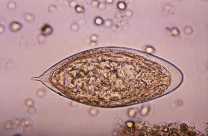
ID#:4843 This micrograph depicts an egg from a Schistosoma haematobium trematode parasite; magnified 500x. Note the egg's posteriorly-protruding, terminal spine, unlike the spinal remnant, which protrudes from the lateral wall of the Schistosoma japonicum egg. These eggs are eliminated in an infected human's feces or urine, and under optimal conditions in a watery environment, the eggs hatch and release "miracidia", which then penetrate a specific snail intermediate host. Once inside the host, the S. haematobium parasite passes through two developmental generations of sporocysts, and are released by the snail into its environment as "cercariae". |
|||
| Tarehe | ||||
| Chanzo | http://phil.cdc.gov/PHIL_Images/20031013/b47fc1793d7443d7a5cdbfbc73d95e53/4843_lores.jpg | |||
| Mwandishi | CDC, Public Health Image Library (PHIL) | |||
| Ruhusa (Kutumia faili tena) |
|
Historia ya faili
Bonyeza tarehe/saa kuona faili kama ilivyoonekana wakati huo.
| Tarehe/Saa | Picha ndogo | Vipimo | Mtumiaji | Maelezo | |
|---|---|---|---|---|---|
| sasa hivi | 17:13, 7 Mei 2006 | | 700 × 460 (38 KB) | Patho | {{Information| |Description=ID#: 4843 Description: This micrograph depicts an egg from the trematode parasite Schistosoma japonicum with its vestigial spine. The Schistosoma japonicum egg is typically oval or subspherical, has a vestigial spine, and is |
Matumizi ya faili
Ukurasa huu umeunganishwa na faili hili:
Matumizi ya faili ulimwenguni
Wiki nyingine hutumia faili hizi:
- Matumizi kwa ar.wikipedia.org
- Matumizi kwa cs.wikipedia.org
- Matumizi kwa de.wikibooks.org
- Matumizi kwa en.wikipedia.org
- Matumizi kwa fr.wikipedia.org
- Matumizi kwa gl.wikipedia.org
- Matumizi kwa ha.wikipedia.org
- Matumizi kwa nl.wikipedia.org
- Matumizi kwa tr.wikipedia.org
- Matumizi kwa zh.wikipedia.org